Stereo-seq for Organoid Research
Spatial Transcriptomics for Organoids
From Development to Therapeutics
Mapping Organoid Biology at Single-Cell Spatial Resolution
Organoids are powerful experimental and preclinical models that mimic the key structural, cellular, and functional features of native tissues. By recreating the complex spatial organization and heterogeneity of real organs, organoids provide unprecedented opportunities to study development, disease progression, and therapeutic response in a controlled yet physiologically relevant context. Stereoseq brings a new dimension to organoid research by combining single-cell resolution with ultra-wide spatial coverage. This enables researchers to map gene expression across the entire 3D architecture of organoids, capturing gradients, niches, and cell–cell interactions that drive organ-level function, accelerating discoveries in developmental biology, regenerative medicine, and drug discovery.
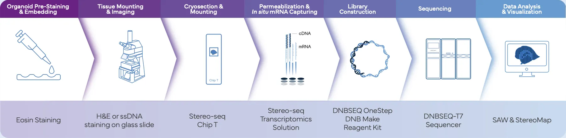
Stereo-seq Workflow For Organoids
Organoids are either labeled with eosin or ssDNA/H&E to visualize structural features, then embedded, sectioned, and mounted on a Stereo-seq chip T for high-fidelity RNA capture. Library construction and sequencing are performed on the DNBSEQ-T7 platform, and data are processed and visualized using SAW and StereoMap software.
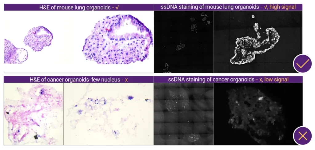
Quality Control Step
The quality control step ensures that only well-preserved and morphologically intact organoids proceed to spatial transcriptomic profiling. High ssDNA signal and clear nuclear morphology indicate suitable samples. This quick morphological assessment helps avoid failed captures and ensures robust RNA profiling in subsequent Stereo-seq steps.
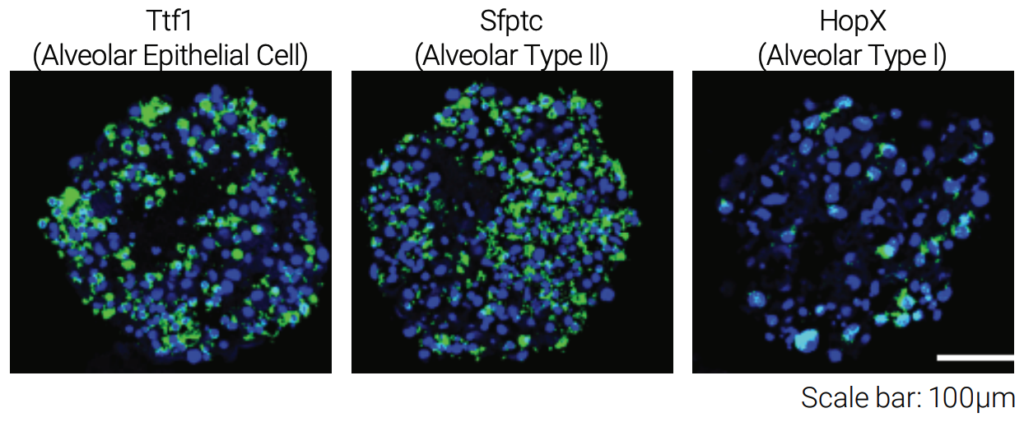
Stereo-seq Revealed Complex, Multicellular Architecture in Organoids
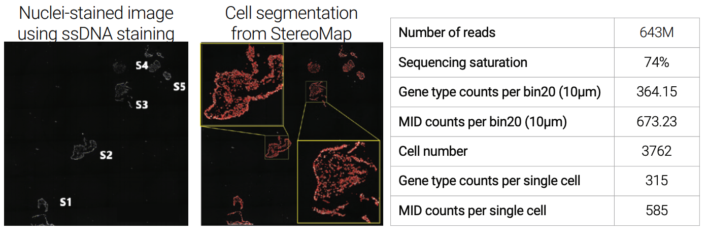
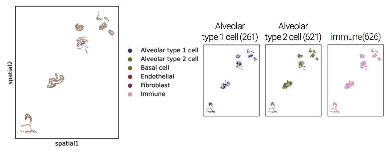
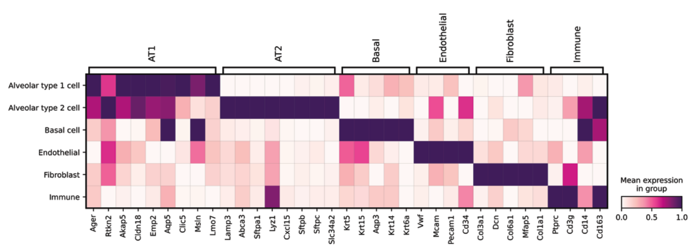
Stereo-seq enables comprehensive spatial profiling of organoids by combining high-resolution imaging and transcriptomic mapping within intact 3D structures. Multiplex immunofluorescence identified distinct epithelial populations (A), and high data quality was confirmed by ssDNA-based nuclear imaging and accurate cell segmentation using StereoMap (B). The architecture within the organoid was revealed by spatially resolving clusters of annotated cell types (C), and corresponding gene expression heatmaps
defined cell–type–specific transcriptional programs (D).
(A)
(B)
(C)
(D)
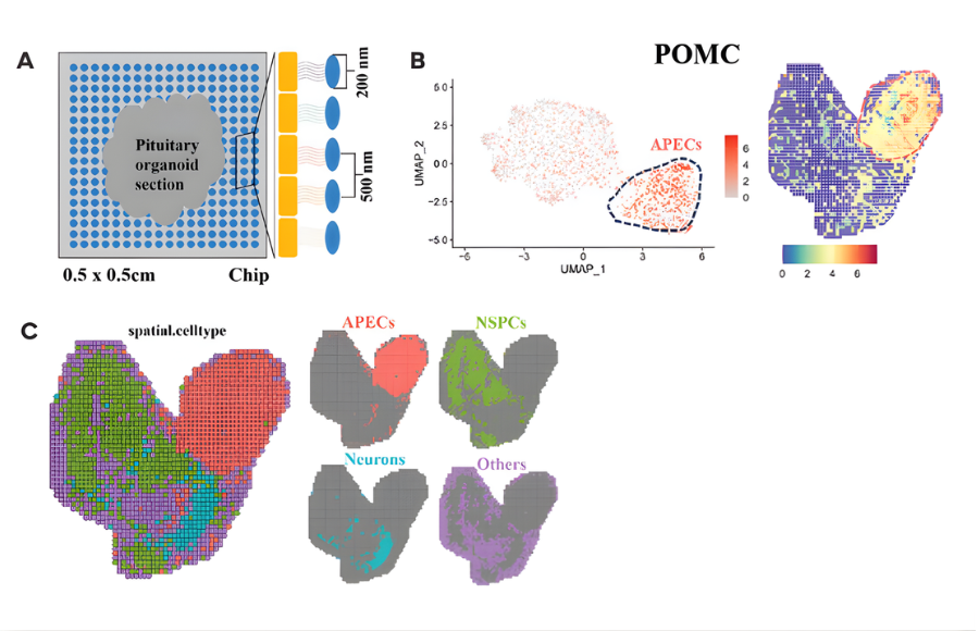
Featured Publication
Human Pituitary Organoids: Transcriptional Landscape Deciphered by Stereo-Seq, with Insights into SOX3’s Role in Pituitary Development
Wang et al., Advanced Science, 2025.