
Whole Transcriptome
& 100+ Proteins
Spatial Proteo-Transcriptomics for Deeper Biological Insight
In Situ Spatial Mapping of RNA & Protein
Stereo-CITE Proteo-Transcriptomics combines unbiased whole-transcriptome RNA profiling with 100+ protein markers from the same fresh-frozen tissue section. By preserving spatial context across both modalities, Stereo-CITE enables deeper investigation of tissue structure, molecular variation, and local cellular interactions.
Benefits of Stereo-CITE Transcriptomics
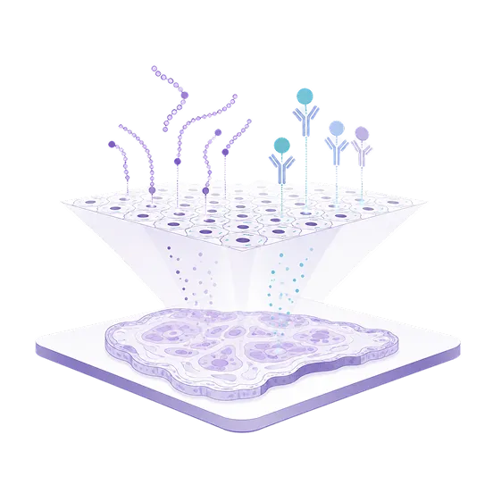
Whole Transcriptome & 100+ Protein Profiling
Capture unbiased whole-transcriptome and high-plex protein expression from the same tissue section.
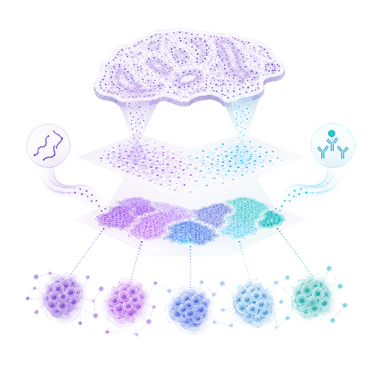
True Single Cell
Resolution
Map cellular organization and tissue architecture in their native spatial context with subcellular resolution.

Reveal Functional Cell Phenotypes
Integrate RNA and protein insights to identify cell states, immune populations, and cellular interactions.
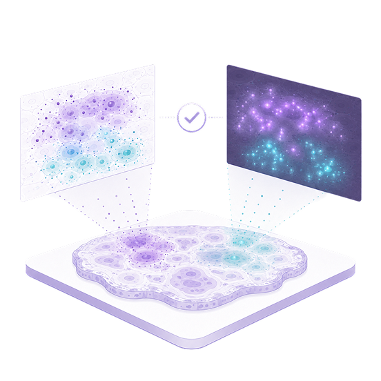
Validated Protein
Specificity
Correlate spatial protein signals with immunofluorescence staining to support confident protein detection.
Why It Matters
See What RNA or Protein Alone May Miss
RNA reveals transcriptional programs, while proteins add complementary information about functional phenotypes and cell-surface identity. Integrating both RNA and protein readouts from the same tissue section can distinguish cell populations and tissue structures that may be less apparent with either modality alone.
HOW IT WORKS
Spatial RNA and Protein Profiling with Stereo-CITE
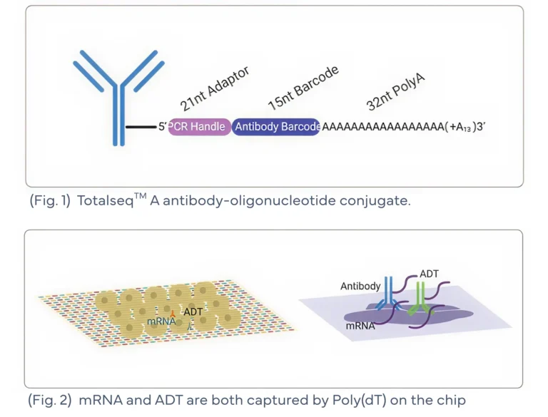
Stereo-CITE combines Stereo-seq with compatible antibody-derived tags (ADTs) chemistry to enable spatial RNA and protein profiling from the same tissue section.
Protein targets are labeled with oligo-conjugated antibodies carrying unique barcodes and poly(A) tails (Fig. 1). During capture, both mRNA and ADTs bind to the chip’s poly(dT) surface and are sequenced together (Fig. 2).
Map Protein Markers in High-Resolution
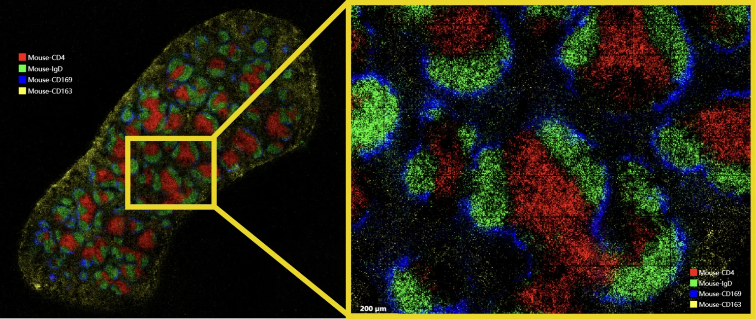
Stereo-CITE spatially maps multiplexed immune protein markers in mouse spleen tissue, including CD4, IgD, CD169, and CD163. The magnified view highlights distinct marker-defined regions, demonstrating how high-resolution protein profiling can resolve immune compartments and local tissue organization in situ.
WORKFLOW
Complete Spatial Transcriptomics and Protein Profiling Workflow
Application
Integrated RNA-Protein Profiling Reveals Human Lymph Node Organization
Stereo-CITE combines whole-transcriptome and high-plex protein profiling to reveal tissue compartments, recapitulate lymph node architecture, and resolve distinct cell states.
01
Multiplexed Protein Mapping Defines Immune and Tissue Compartments
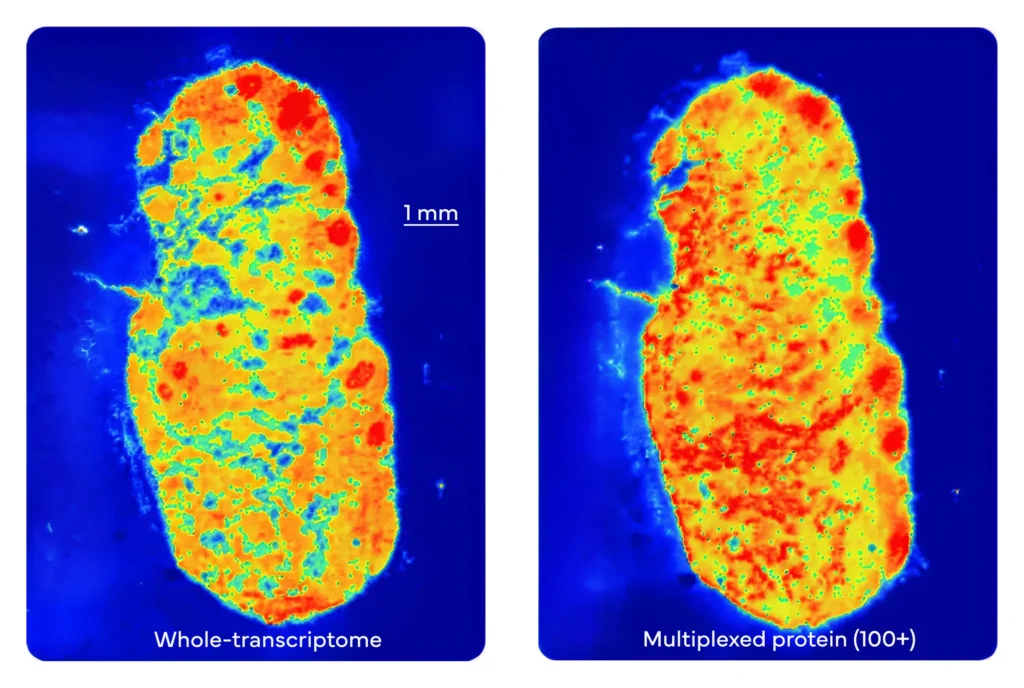
RNA & 100+ Protein Markers
Stereo-CITE proteo-transcriptomics solution co-profiles whole-transcriptome RNA and 100+ protein markers in human paracancer lymph node tissue (Bin20).
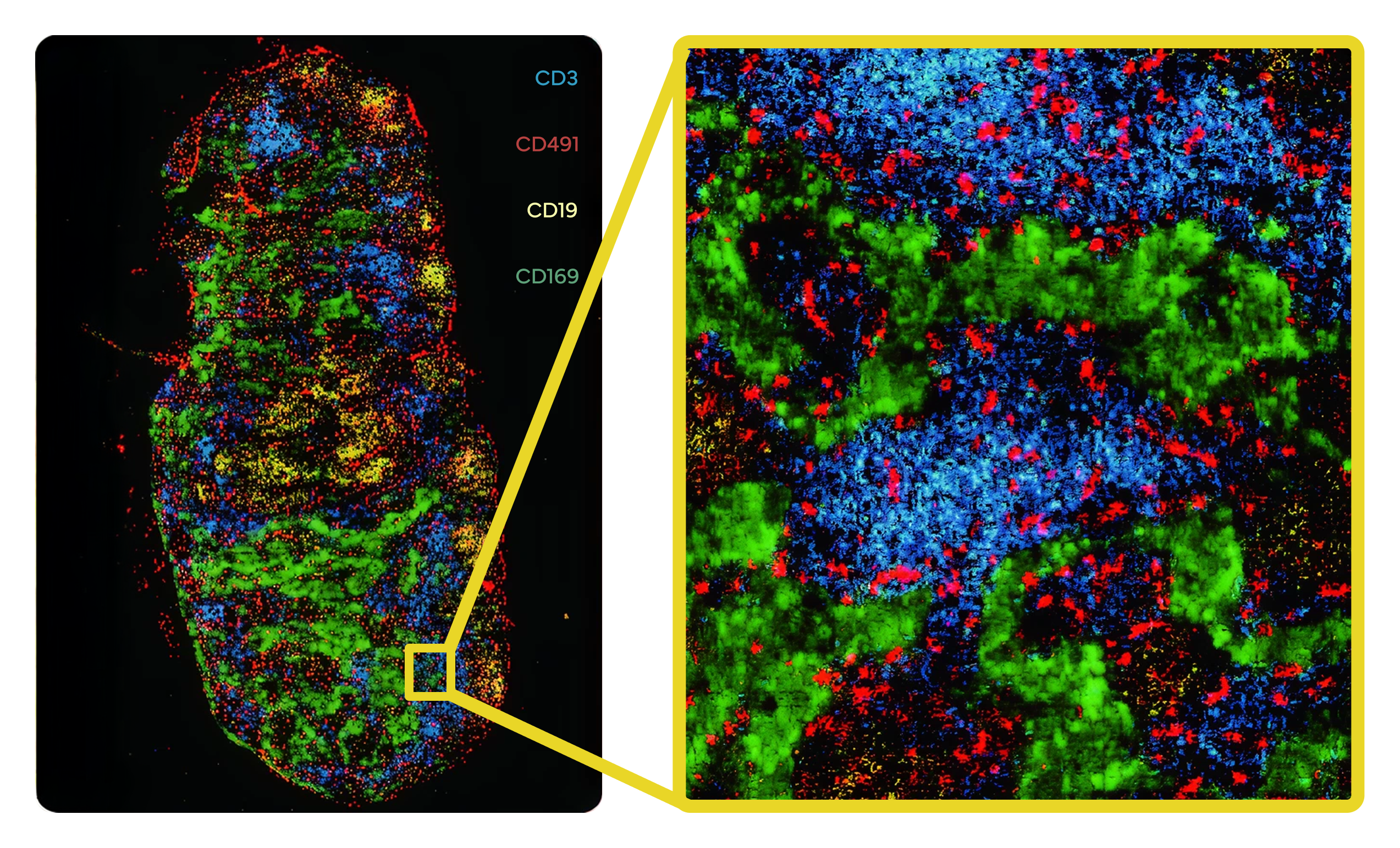
Marker-Based Tissue Mapping
Selected markers with pseudo-colors, including CD3, CD49f, CD19, and CD169, highlight distinct tissue compartments and connect spatial gene expression with protein-defined cellular phenotypes.
02
Spatial Co-Clustering Recapitulates Tissue Architecture
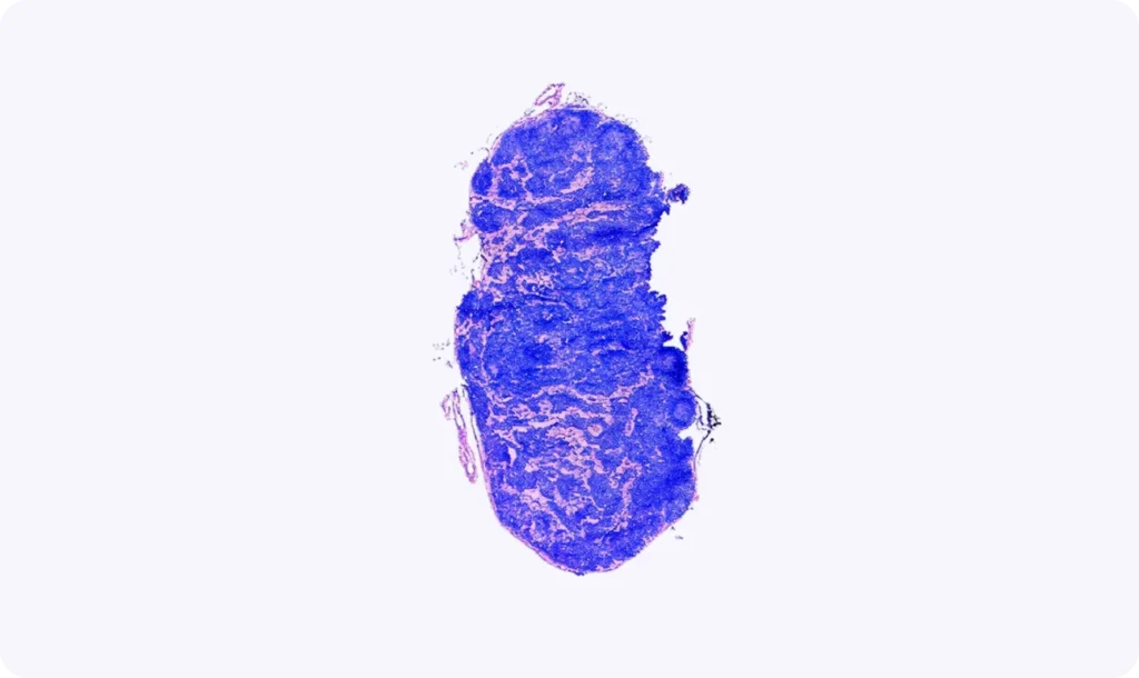
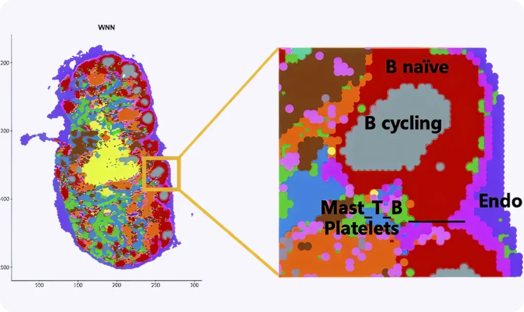
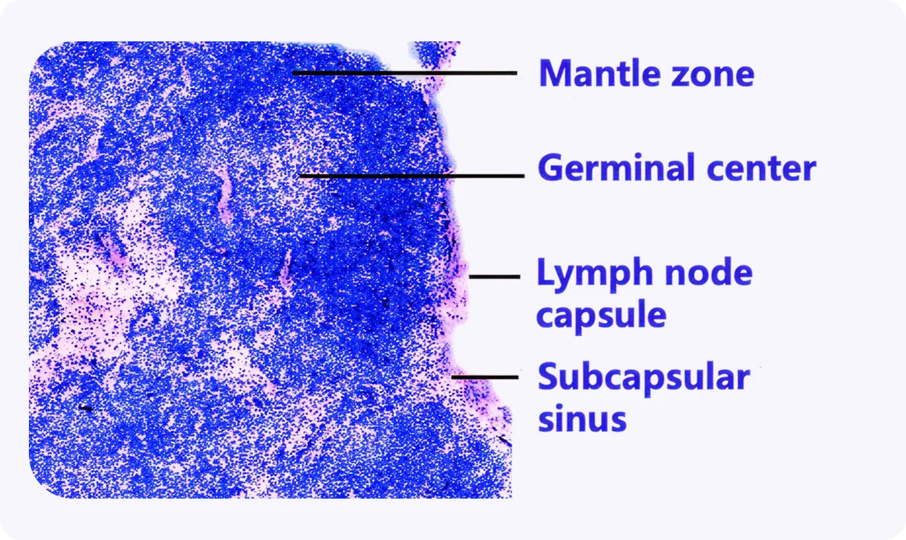
Spatial co-clustering builds on the co-profiled RNA-protein readout to recapitulate lymph node tissue architecture observed by H&E staining, including the mantle zone, germinal center, lymph node capsule, and subcapsular sinus.
H&E Section Staining
H&E staining of adjacent section
Spatial Co-Clustering
Spatial co-clustering results of human paracancer lymph nodes
Pathology Annotations
H&E staining with pathology annotations of cancer
03
Integrated RNA-Protein Clustering Improves Cell-State Resolution
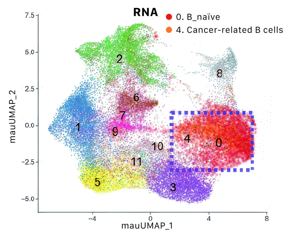
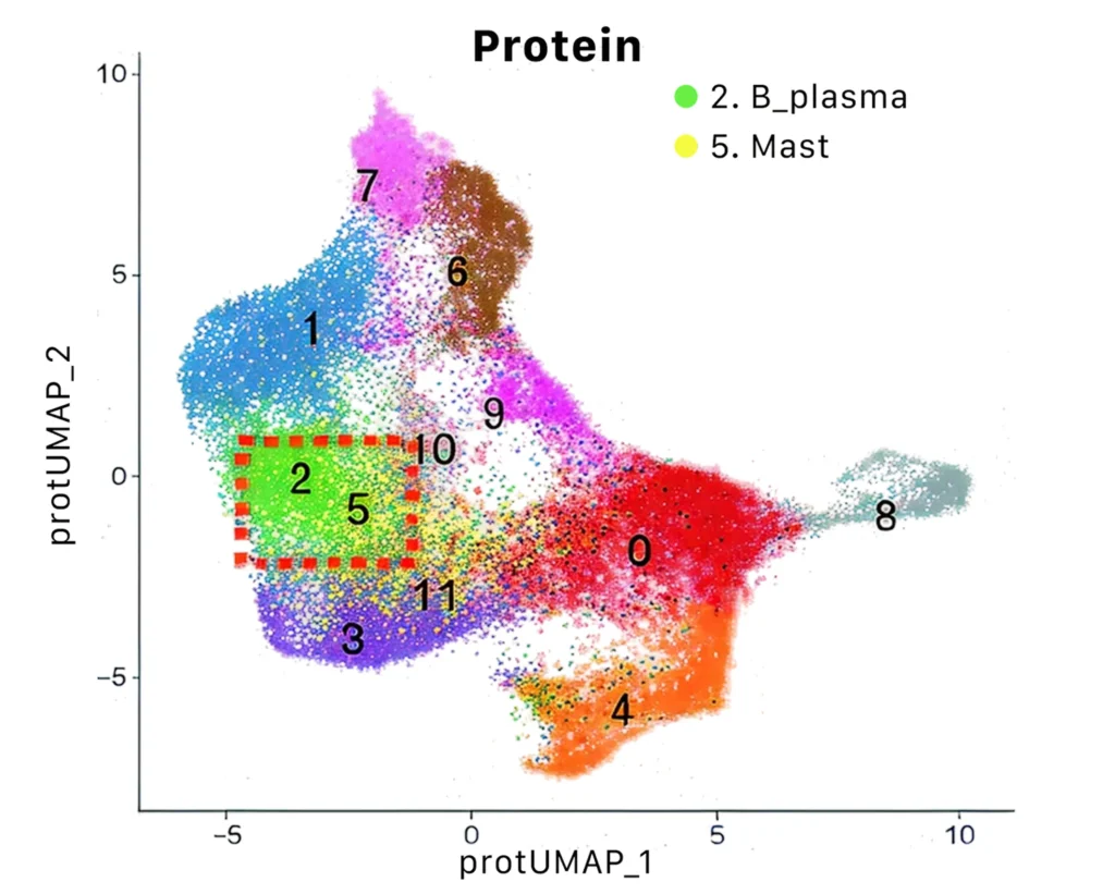
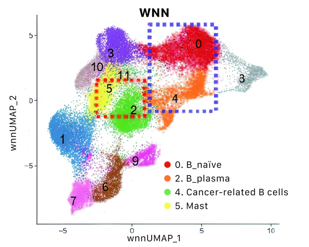
Integrated RNA-protein clustering further distinguishes cell populations that are less clearly separated by RNA-only or protein-only analysis, supporting improved resolution of B cell subsets, endothelial cells, mast cells, and cancer-associated populations.
04
RNA and Protein Reveal Complementary Cell-State Information
The modality weight analysis shows that RNA and protein signals contribute differently across cell states, highlighting the complementary value of each readout in integrated clustering. Protein information contributes strongly to specific immune and tissue-associated populations, while RNA remains important for transcriptome-defined cell identity. Together, these weighted inputs help improve cell-state resolution beyond RNA-only or protein-only analysis.
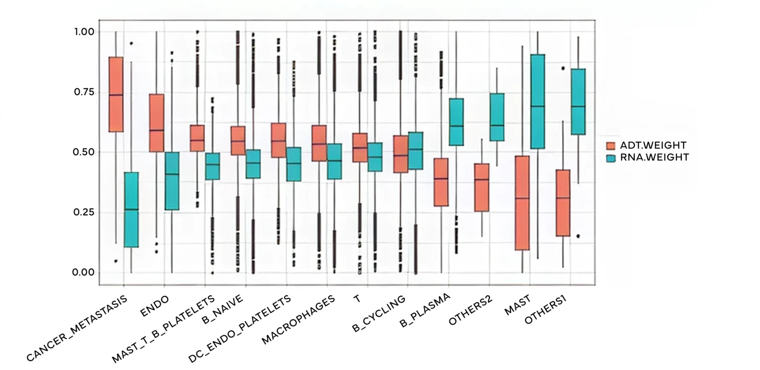
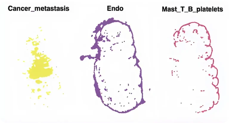
Protein-Enriched Clusters
Protein information contributed strongly to resolving select cell populations, including cancer metastasis-associated cells, endothelial cells, and mixed immune or platelet-associated groups. These results highlight how protein markers can sharpen spatial cell-state definitions that may be less distinct from transcriptomic data alone.
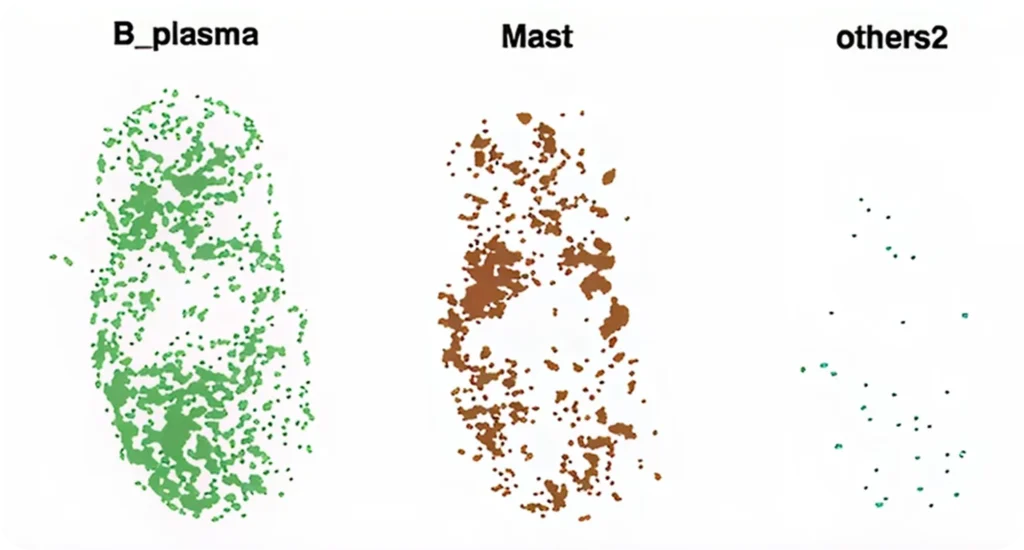
Transcriptome-Enriched Clusters
Transcriptome information contributed more strongly to clusters such as B plasma cells, mast cells, and additional cell populations. This supports the value of whole-transcriptome profiling for defining gene-expression-based cell identities within the tissue.
How Stereo-CITE Stands Out

30,000+ genes
Unbiased Whole-transcriptome profiling enables broad, unbiased spatial RNA analysis beyond targeted gene panels.

No automation
Run a manual-friendly workflow without requiring a dedicated automation system.

100+ proteins
Profile high-plex protein expression together with spatial transcriptome data from the same tissue section.

20 samples/week
Designed to support practical study throughput with 10–20 samples per week using 1–2 FTEs.
publications
Spatial Transcriptomics and Protein Profiling Publications
Explore Stereo-CITE publications demonstrating spatial transcriptomics and protein profiling from the same tissue section, combining whole-transcriptome analysis with high-plex protein-level phenotyping in a single workflow.
Demo Data
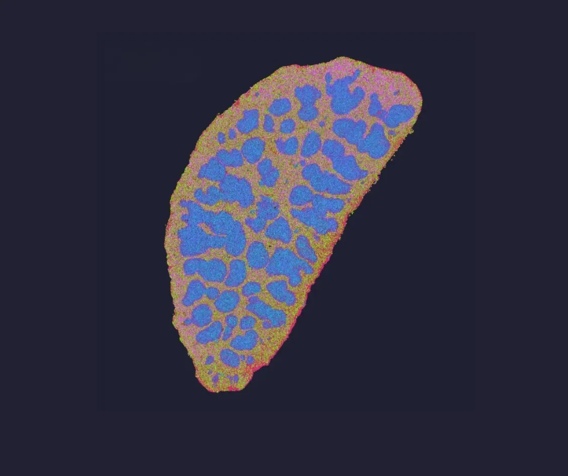
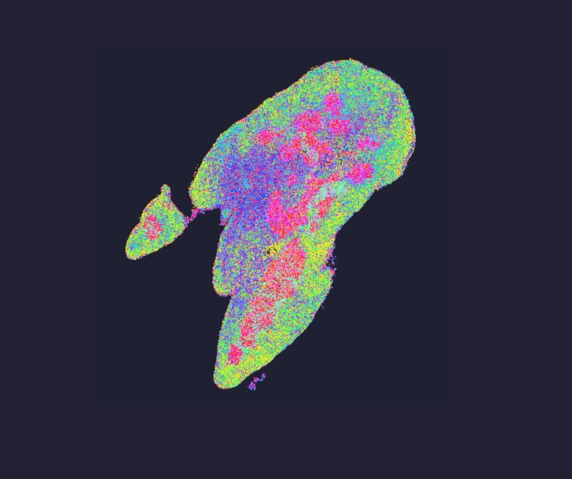
Explore The Power of Stereo-CITE with our Demo Datasets
Experience interactive spatial data visualization by simply clicking on each image below.
Ordering Information
| Category | Product | Cat. No. |
|---|---|---|
| Sample Prep | Stereo-CITE Proteo-Transcriptomics Set V1.1 | 211PT11114-CG |
| Stereo-seq Protein Assisted Kit (included in the CITE kit) | 212KA11114-CG | |
| Permeabilization Sets | Stereo-seq Permeabilization Set for Chip-on-slide | 211SP11118-CG |
| Library Prep | Stereo-seq 16 Barcode Library Preparation Kit V1.1 | 111KL11160-CG |